
Instrumentation and software systems for analysis of the microcirculation
|Welcome| - |CapiScope CAM1 Capillary Anemometer| - |CapiScope HVCS Video Capillaroscopy| - |Sample Images and Videos| - |User Guides| - |References| - |Customers| - |Downloads| - |Contacts|
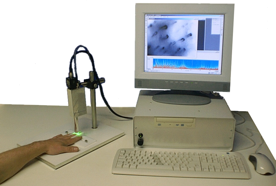
CapiScope Capillaroscopy System CAM1 Laser Doppler Capillary Anemometer
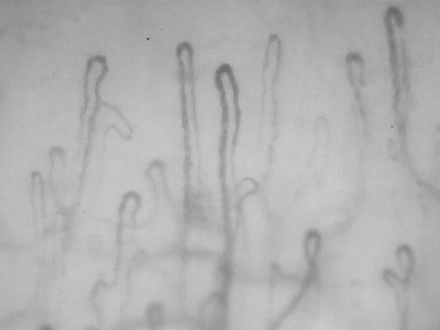
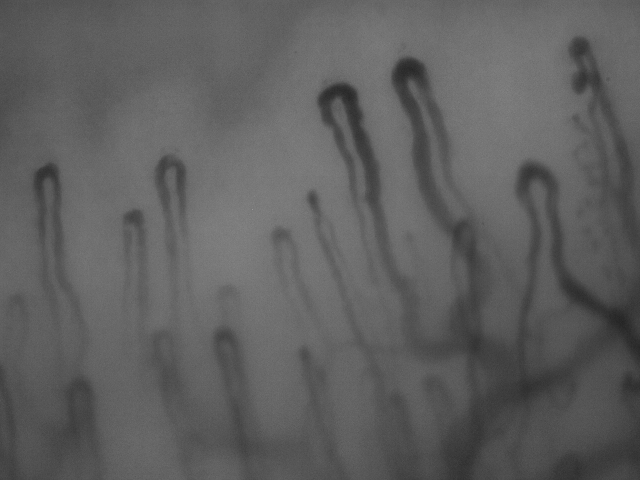
Measurement of Capillary Morphology and Capillary blood cell velocity
- Visualize, capture and record capillaries digitally.
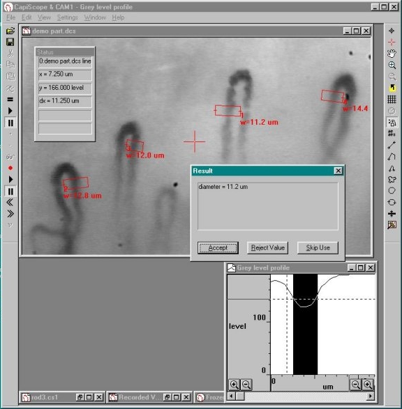
- Automatic measurement of vessel diameters.
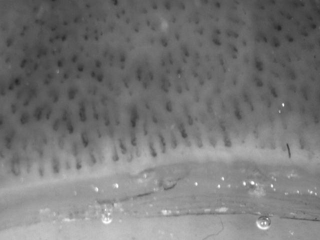
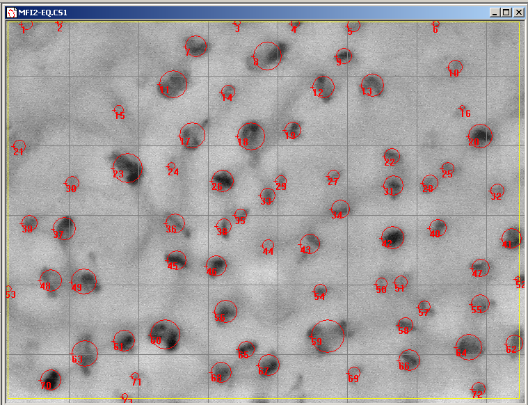
- Manual and automatic capillary density.
- Blood cell velocities from 0.1 mm/s to 15 mm/s using the internal laser.
- Blood cell velocity from 0 to about 2mm/s using video correlation.
- Fast response time follows cardiac and vasomotion velocity changes.
- Laser velocity measurements are NOT limited to nailfold capillaries.
- High quality video capillaroscopy images.
- Velocity can be measured from tissues with poor image quality using the laser.
- New small size and light weight for easier positioning.
Video and Image Recording
CapiScope can display and record video at full frame and full frame rate. The monochrome video is stored uncompressed to avoid problems with information loss found with many digital video recorders. Single frames can be saved in bmp or tiff formats and video can be exported as avi files.
Capillary blood cell velocity by laser Doppler
The CAM1 Capillary Anemometer measures blood cell velocity using an internal low power near infrared (780nm) laser. This is focused via the objective down to a 10 micron diameter spot in the centre of the field of view. The CAM1 laser can be positioned onto the apex of a capillary loop using the XYZ micropostioners.
The laser beam is reflected by blood cells at the focal point moving perpendicular to the tissue surface. This gives the laser light a Doppler shift directly proportional to the velocity of the reflecting blood cells. The Doppler shifted laser is collected by the objective and internal optoelectronics, digitised by the CAMDAQ USB module and processed by the CapiScope laptop computer. The Doppler shift is processed in real time to produce a velocity trace in absolute units of mm/s.
The velocity trace can be saved together with an image or video sequence of the capillary being measured onto the hard disc of the CapiScope laptop.
Due to the huge number of AI bots repeatedly downloading everything, our videos now ask for a username and password. Please use "user" and "capiscope".
Capillary blood cell velocity by video correlation
Velocity can also be measured from live video, video recorded digitally by the CapiScope capture and analysis program, avi files, or video from a VHS or digital video recorder*.
Velocity is measured by using the mouse to draw a line along the vessel. The vessel can have any orientation, and doesn't even need to be straight. The line thickness can be adjusted so that it covers the width of the capillary. This gives an average of the pattern over the whole width.
The grey level profile along the line is taken for each video frame every 1/25th of a second. Faster frame rates upto 600 fps are possible by using a smaller region of the image.
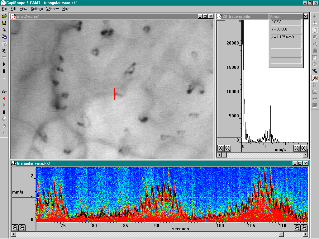
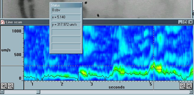
The grey level pattern along each line is compared to the pattern from the previous frame (or, optionally, several frames before for very low velocities). The comparison is performed by calculating the correlation coefficient for every possible shift of the previous grey level profile relative to the new profile. The shift which produces the highest correlation indicates the distance travelled between the two grey level profile measurements, and hence the velocity. CapiScope displays the correlation, along with the velocity trace (cbv) as a colour map, showing red for a high correlation through to blue for low correlation. The cbv (shown as a black trace) can be averaged over any period, and the table of averaged results can be saved or easily pasted into other applications.
SPECIFICATIONS
Specifications
Anemometer
Velocity ranges: 1.8, 3.7, 7.3, & 14.6 mm/s
Resolution: 8 bits
Velocity measurement range: 0.05 mm/s to 14.6 mm/s (max. Doppler shift 50 kHz).
Laser power: 1 mW typical, 1.5 mW max
Laser wavelength: 780 nm typical.
Measurement area: 8 um x 16 um approx.
Imaging
| CAM1 | CAM1-HR3 | |
| Image size (pixels) | 1280 x 1024 | 2048 x 1536 |
| standard lens: | ||
| Magnification (μm/pixel) | 0.81 | 0.546 |
| field of view (μm) | 1037 x 829 | 1118 x 839 |
| low power lens: | ||
| Magnification (μm/pixel) | 1.71 | 1.14 |
| field of view (μm) | 2186 x 1749 | 2333 x 1750 |
Illumination: 4 x 525 nm light sources.
Video output: USB uncompressed.
Dimensions
CAM1:
235 x 90 x 120 mm including lens and xyz.
weight 750g.
235 x 30 x 60 mm including lens.
weight 400g.
CAMDAQ Interface Module
86 x 57 x 26 mm
weight 100g
Operating Conditions
Temperature +10oC to +35oC
Relative Humidity 30% to 75% noncondensing
Altitude 700 to 1060 hPa
Shipping and Storage Conditions
Temperature -10:sup:oC to +45oC
Relative Humidity 95% noncondensing maximum
Software
CapiScope software requires Windows XP, Vista, 7, 8, 10 or 11.
Standards
Note
This is not a medical device, it has no diagnostic nor therapeutic function. For research use only (except in the UK excluding NI).
EN 60601-1:2006 /AC:2010 /A1:2013 Medical electrical equipment -- Part 1: General requirements for basic safety and essential performance
EN 60601-1-2:2007 Medical electrical equipment -- Part 1-2: General requirements for basic safety and essential performance - Collateral standard: Electromagnetic compatibility - Requirements and tests
EN ISO 14971:2012 Medical devices - Application of risk management to medical devices.
EN 60825-1:2007 Safety of laser products. CLASS 1.
UKCA UK MDR 2002 (SI 2002 No. 618) [ Class 1 non-sterile ]
All devices prior to 2021-01-01: CE Medical Devices Directive 93/42/EEC Annex VII. [ class I device non-sterile ].
Here is a very old quick demonstration video using the CAM1:
Due to the huge number of AI bots repeatedly downloading everything, our videos now ask for a username and password. Please use "user" and "capiscope".
|Welcome| - |CapiScope CAM1 Capillary Anemometer| - |CapiScope HVCS Video Capillaroscopy| - |Sample Images and Videos| - |User Guides| - |References| - |Customers| - |Downloads| - |Contacts|
Copyright 2024 KK Technology
KK Research Technology Ltd, Honiton, Devon, EX14 1YL, England, United Kingdom, +44 140446242